Human Heart
The four-chambered muscular pump at the centre of the circulatory system — its anatomy, its beat, and the medicine built around it.
The human heart is a muscular organ about the size of a fist that sits in the mediastinum between the lungs and drives blood through the pulmonary and systemic circuits, beating some 100,000 times a day. Four chambers, four valves and a coronary circulation of its own keep oxygen moving to every tissue, while an electrical conduction system beginning at the sinoatrial node sets the rhythm of each beat. Cardiovascular disease remains the leading cause of death worldwide — an estimated 19.8 million deaths in 2022 — and the heart's modern history runs from William Harvey's account of the circulation in 1628 to the first human heart transplant in 1967.
In this article
- 1Overview
- 2Location and External Form
- 3Dimensions and Mass
- 4Chambers and Septa
- 5Valves and the Direction of Flow
- 6Wall Structure and Tissue
- 7Coronary Circulation
- 8Electrical Activity and the Cardiac Cycle
- 9Pumping Performance
- 10Regulation of Stroke Volume
- 11Daily Workload
- 12Disease Burden
- 13The Heart in Microgravity
- 14Milestones in Heart Science and Surgery
- 15Key Figures
- 16Frequently Asked Questions
- 17Timeline: from Harvey's circulation to the modern transplant era
- 18Knowledge graph
Overview
The human heart is a fist-sized muscular organ that pumps blood throughout the body. It sits at the center of the circulatory system, a network of arteries, veins and capillaries that carries blood to and from every area of the body. Each beat is a muscular contraction that pushes blood to the lungs and to the rest of the body.
1. Location and External Form
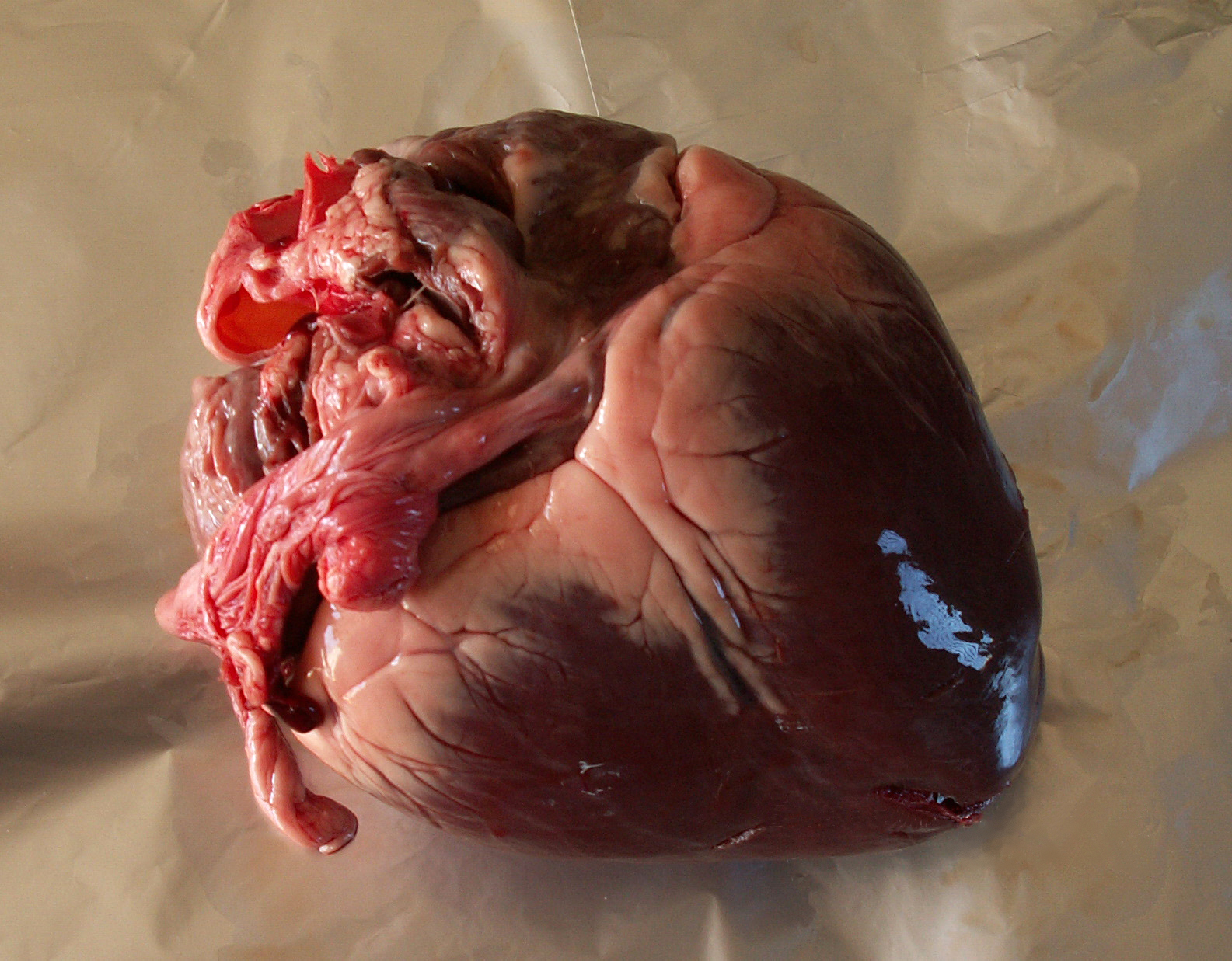
The heart occupies the mediastinum, the central region of the thoracic cavity, where it sits between the lungs and behind the sternum. Roughly two-thirds of the organ lies to the left of the midline.
In form the heart resembles a pinecone: broad across its upper surface and narrowing toward the tip. Five surfaces are distinguished — a posterior base, an inferior diaphragmatic surface, an anterior sternocostal surface, and left and right pulmonary surfaces — together with right, left, superior and inferior margins. The superior and inferior venae cavae, the aorta and the pulmonary trunk all attach to the upper surface, the region known as the base. Grooves on the external surface carry branches of the right and left coronary arteries, the vessels that deliver blood to the heart muscle.
2. Dimensions and Mass
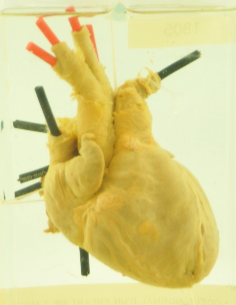
The measurements below collect the principal dimensions and mass of the adult heart3Anatomy and Physiology 2e, 19.1 Heart Anatomy · OpenStax, Rice University ↗.
3. Chambers and Septa
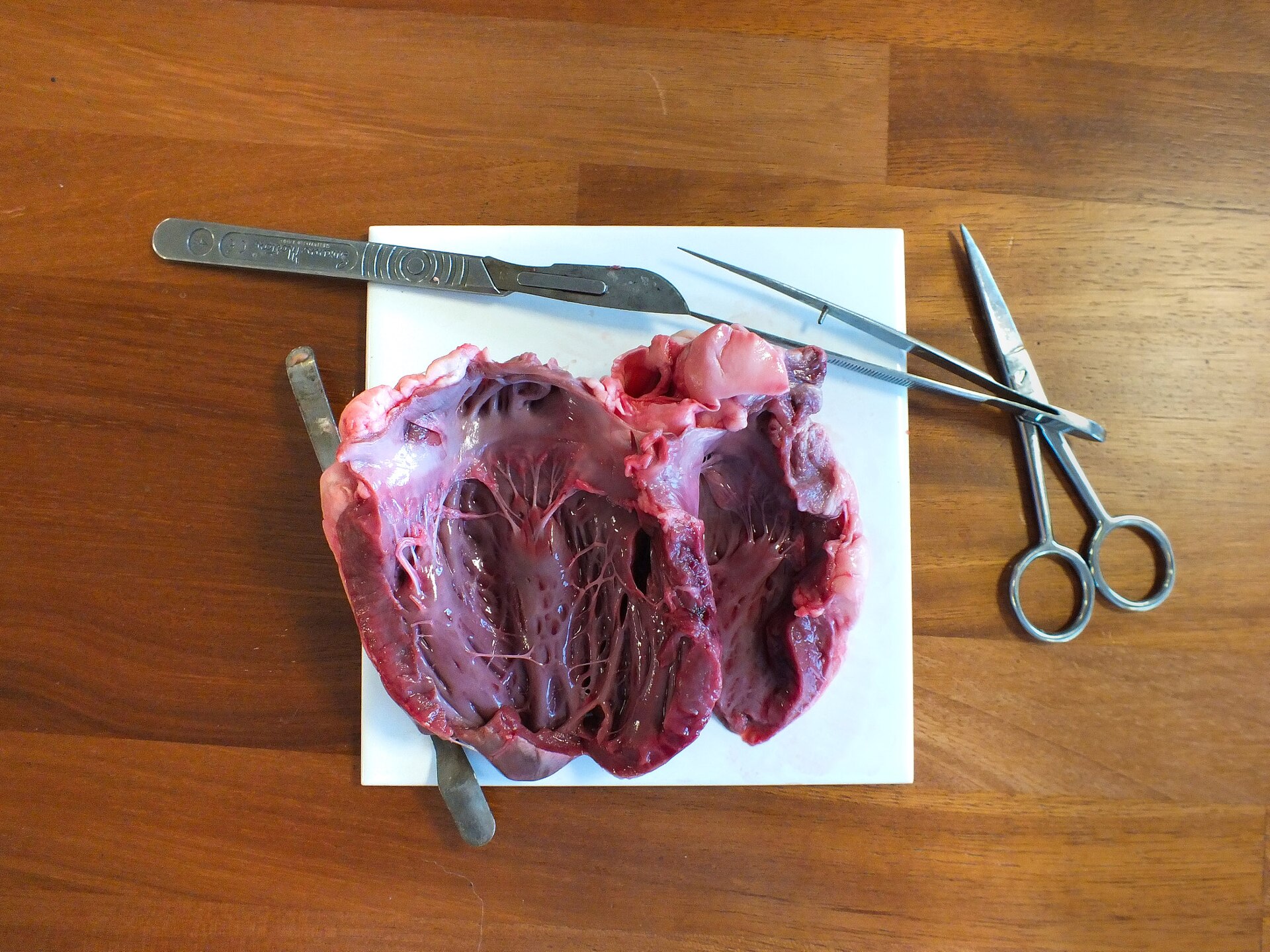
The heart is built around four chambers: two upper atria that receive blood and two lower ventricles that pump it out. The atria push blood into the ventricles, which then send it onward to the lungs or to the body. Together the two sides form a right and a left pump, serving the pulmonary and the systemic circuits.
A septum divides the right side of the heart from the left, separating the chambers into the interatrial and interventricular septa. Blood passes between chambers through the atrioventricular orifices. The left ventricle is the largest and strongest chamber, its wall much thicker than that of the right, although both sides pump the same volume.
4. Valves and the Direction of Flow
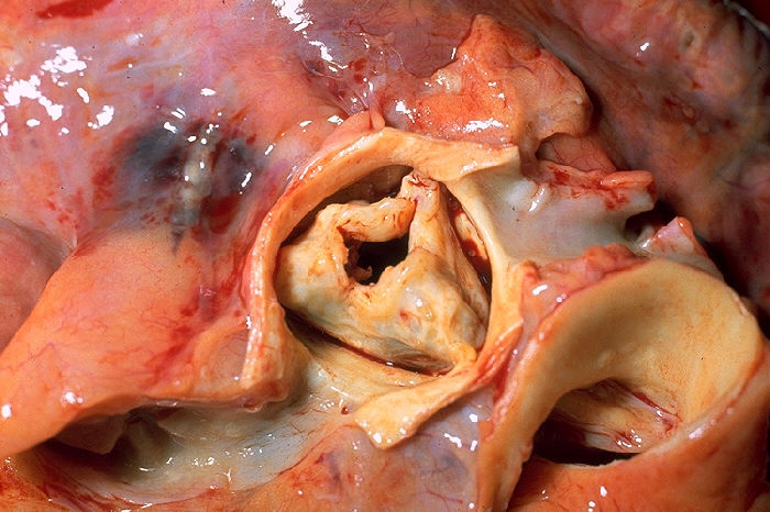
Four valves regulate the flow of blood through the heart, and each of them is placed so that blood can move in only one direction. The tricuspid valve sits between the right atrium and the right ventricle, while the mitral valve occupies the same position on the left, between the left atrium and the left ventricle. Blood leaving the ventricles passes through two further valves: the pulmonary valve, which carries blood from the right ventricle to the lungs, and the aortic valve, which opens from the left ventricle into the aorta. The atrioventricular valves — those guarding the entrances to the ventricles — prevent blood from flowing back from the ventricles into the atria.
The two atrioventricular valves are not identical in construction. The tricuspid valve on the right side has three cusps, whereas the left valve is known as the mitral, or bicuspid, valve because it has only two. This arrangement gives each ventricle an inlet valve from its atrium and an outlet valve to its artery, so that the course of blood through the heart is held to a single direction.

5. Wall Structure and Tissue
The heart wall is built from three layers: a thin inner endocardium, a thick muscular myocardium, and an outer pericardium sac. The pericardium is a serous sac of two layers, and its strong fibrous outer portion is attached to the diaphragm, the mediastinal pleura and the sternum. A small amount of fluid between the layers of the pericardium lowers friction as the beating heart moves against nearby tissues.
Cardiac muscle is built from contractile cells, which form the bulk of atrial and ventricular tissue, and conducting cells, which make up the conduction system. Neighbouring cardiac muscle cells bind together at intercalated discs, structures built from desmosomes and many gap junctions that let ions move between the cells and support a synchronized contraction. The muscle works aerobically and keeps myoglobin, lipids and glycogen stored in its cytoplasm. Its cells contract in single twitches with long refractory periods and brief periods of relaxation that let the heart refill.
6. Coronary Circulation
The heart muscle is nourished by its own vascular supply, the coronary arteries, which arise from the first part of the aorta just above the aortic semilunar valve and divide into the left and right coronary arteries. Grooves visible on the heart's external surface carry branches of these right and left vessels, conducting blood to the muscle they serve. Of the two main channels, the left main artery carries 80% of the flow to the heart muscle.
7. Electrical Activity and the Cardiac Cycle
The adult heart can generate its own electrical impulse, its rate set by whichever of its cells fires fastest, and the signal spreads rapidly from cell to cell. The components of this conduction system are the sinoatrial node, the atrioventricular node, the atrioventricular bundle, its branches, and the Purkinje cells. The nervous and endocrine systems modulate that intrinsic rate rather than originating it. Each heartbeat begins with pacemaker cells in the sinoatrial node of the right atrium — the heart's main pacemaker, which sits at the junction of the superior vena cava and the right atrium and discharges about 70 times a minute; on reaching the atrioventricular node the signal slows briefly while the ventricles fill, then travels from the top of the heart to the bottom to make it contract.
8. Pumping Performance
Ejection fraction expresses the share of the end diastolic volume that the heart pumps out with each contraction; it is found by dividing stroke volume by the end diastolic volume and is normally reported as a percentage.11Anatomy and Physiology 2e, 19.4 Cardiac Physiology · OpenStax, Rice University ↗
Cardiac output measures how much blood one ventricle pumps in a minute, and it is obtained by multiplying the stroke volume by the heart rate.11
In a healthy adult the stroke volume usually falls between about 55 and 100 millilitres and the resting heart rate is around 75 beats per minute, which gives a cardiac output near 5 litres per minute.11
| Measure | Value |
|---|---|
| Resting heart rate | around 75 beats per minute11 |
| Stroke volume | about 55 to 100 millilitres11 |
| Cardiac output | near 5 litres per minute at rest, with a mean of about 5.25 litres per minute and a range of 4.0 to 8.0 litres11 |
| End diastolic volume | about 130 millilitres of blood in a resting adult, just before the ventricles contract12Anatomy and Physiology 2e, 19.3 Cardiac Cycle · OpenStax, Rice University ↗ |
| End systolic volume | about 50 to 60 millilitres of blood left in the ventricle after contraction12 |
| Ejection fraction | 55 to 70 percent of the end diastolic volume, with a mean of about 58 percent11 |
9. Regulation of Stroke Volume
Stroke volume is governed mainly by three factors: the preload, or stretch on the ventricles before they contract; the contractility, or force of the contraction itself; and the afterload, or pressure the ventricles must overcome. Under the Frank-Starling mechanism, stretching the ventricular muscle within its physiological limits makes it contract more forcefully, so a larger filling volume produces a larger stroke volume.
10. Daily Workload
The average heart beats about 100,000 times a day and pumps about 2,000 gallons (7,571 litres) of blood.
11. Disease Burden
The behavioural risk factors that matter most for heart disease and stroke are an unhealthy diet, physical inactivity, tobacco use and the harmful use of alcohol, while air pollution stands out as an important environmental factor. Among the everyday habits that raise a person's risk are diets high in saturated fat, refined carbohydrates and salt, along with physical inactivity, heavy drinking, smoking and stress. Cutting the burden of cardiovascular disease depends on embedding management interventions in universal health coverage packages, yet many countries still require significant investment in their health systems.
An estimated 19.8 million deaths from cardiovascular diseases occurred worldwide in 2022 — approximately 32 percent of all deaths.
12. The Heart in Microgravity
In weightlessness, blood and other bodily fluids that gravity normally pulls downward shift toward the head, so the cardiovascular system works less to keep blood flowing to the brain. Adaptation to weightlessness can reduce blood volume and the function of the heart and blood vessels. Back on Earth, astronauts may feel weak, dizzy, or faint when standing.
The Engineered Heart Tissues study used 3D heart tissue grown from stem cells to examine cardiac function in microgravity. A magnetic sensor beneath the culture chamber tracked the tissue in real time.
13. Milestones in Heart Science and Surgery
-
1628De Motu Cordis publishedWilliam Harvey's work on the circulation of blood was dedicated to the king, following his recognition that blood circulates fully through the body, supported by experiments and arguments and aided by access to the royal herd of deer and the presentation of medical cases to the king.
-
1893First open heart surgeryDaniel Hale Williams performed the world's first open heart surgery at Provident Hospital, the Chicago institution he founded as the first black owned and operated hospital in the United States, and became chief surgeon at Freedman's Hospital in Washington, D.C. the same year. 16Daniel Hale Williams, M.D. (Opening Doors: Contemporary African American Academic Surgeons) · U.S. National Library of Medicine ↗
-
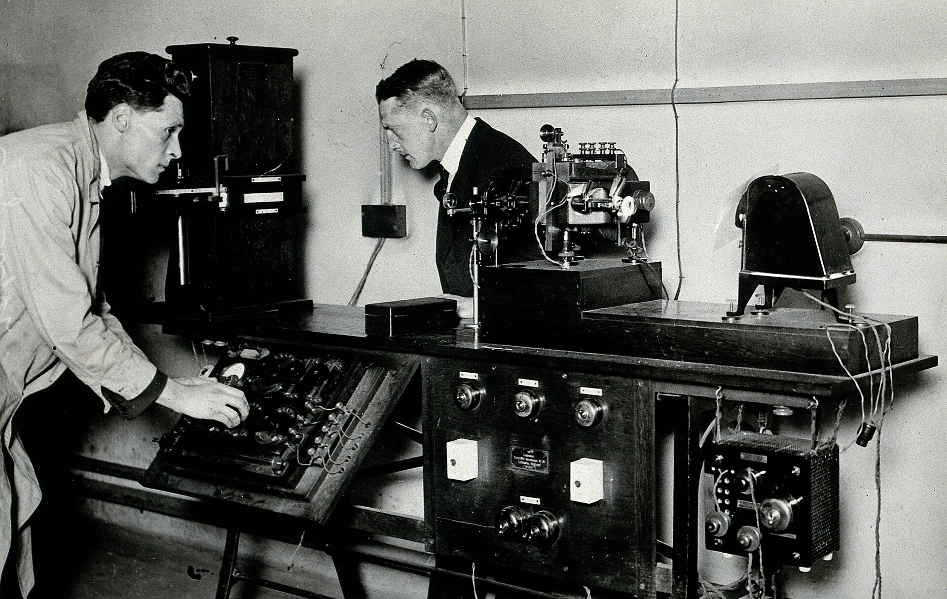
1924Nobel Prize for the electrocardiogramThe Nobel Prize in Physiology or Medicine was awarded solely to Willem Einthoven for discovering the mechanism of the electrocardiogram.
-
1956Nobel Prize for heart catheterizationThe Nobel Prize in Physiology or Medicine was divided equally between Andre Frederic Cournand, Werner Forssmann and Dickinson Richards for discoveries concerning heart catheterization and pathological changes in the circulatory system. 17The Nobel Prize in Physiology or Medicine 1956: Summary · Nobel Prize Outreach ↗
-
December 1967First human heart transplantChristiaan Barnard led twenty surgeons in replacing the heart of the incurably ill South African grocer Louis Washkansky with a heart from a fatally injured accident victim; the transplant succeeded, though Washkansky died eighteen days later from double pneumonia after anti-rejection drugs destroyed his immunity. 18Christiaan Barnard · Encyclopaedia Britannica ↗
14. Key Figures
The English physician William Harvey was the first to recognize that blood circulates fully through the human body, and he backed the idea with experiments and arguments19William Harvey · Encyclopaedia Britannica ↗.
Daniel Hale Williams, who lived from 1856 to 1931, was a leading surgeon and educator and the founder of Provident Hospital and Training School in Chicago, the first black owned and operated hospital in the United States16.
Einthoven was the sole laureate of the 1924 Nobel Prize in Physiology or Medicine, and the citation credits his discovery of the mechanism of the electrocardiogram20The Nobel Prize in Physiology or Medicine 1924: Summary · Nobel Prize Outreach ↗.
15. Frequently Asked Questions
How many chambers and valves does the heart have?
The heart has four chambers and four valves. The four valves are the tricuspid, mitral, pulmonary, and aortic valves 7How the Heart Works: How Blood Flows through the Heart · National Heart, Lung, and Blood Institute (NIH) ↗.
What is the difference between the pulmonary and systemic circuits?
The heart's four chambers are organized into two pumps, a right one and a left one, providing blood flow to the pulmonary and systemic circulations 10Anatomy, Thorax, Heart (StatPearls) · StatPearls Publishing / NCBI Bookshelf ↗.
Where does each valve sit?
How big and how heavy is the human heart?
What is the normal resting heart rate, and how much blood does each beat move?
How much blood sits in a ventricle before and after it contracts?
What is a normal ejection fraction?
It falls between 55 and 70 percent, with a mean of about 58 percent 11.
What do the two heart sounds come from?
How common is heart disease worldwide?
Cardiovascular diseases caused an estimated 19.8 million deaths in 2022, approximately 32 percent of all deaths worldwide 13Cardiovascular diseases (CVDs) · World Health Organization ↗. Heart disease is the leading cause of death in the United States 14Heart Diseases · MedlinePlus, U.S. National Library of Medicine ↗.
16. Timeline: from Harvey's circulation to the modern transplant era
- 1 April 1578William Harvey is bornHarvey was born in Folkestone, Kent, and became the English physician who first recognised the full circulation of the blood in the human body.
- 25 April 1602Harvey earns his doctorate at PaduaHe received his doctorate from the University of Padua and returned to England to work as a doctor.
- 1615Appointed Lumleian lecturerHarvey became Lumleian lecturer in surgery at the Royal College of Physicians of London, a post he held for more than forty years.
- 1618Physician to the kingHarvey was appointed physician to James I and continued as physician to Charles I after his accession in 1625.
- 1628De Motu Cordis is publishedHarvey's most important book set out his account of the circulation of the blood and was dedicated to the king.
- 3 June 1657Death of William HarveyHarvey died in London, roughly half a century before the first measurements of blood pressure and more than two centuries before the heart could be operated on.
- 1856Daniel Hale Williams is bornWilliams became a leading surgeon, educator and founder of Provident Hospital and Training School in Chicago, the first black owned and operated hospital in the United States.
- 1893The world's first open heart surgeryWilliams performed the world's first open heart surgery at Provident Hospital in Chicago, and in the same year was appointed chief surgeon at Freedman's Hospital in Washington, D.C.
- 1898Williams returns to ProvidentAfter serving as professor of surgery at the Howard University Medical Department, he returned to Provident Hospital in Chicago.
- 1924Nobel Prize for the electrocardiogramWillem Einthoven received the Nobel Prize in Physiology or Medicine for his discovery of the mechanism of the electrocardiogram.
- 1956Nobel Prize for heart catheterizationAndré Cournand, Werner Forssmann and Dickinson Richards shared the prize for their discoveries concerning heart catheterization and pathological changes in the circulatory system.
- 3 December 1967First human heart transplantChristiaan Barnard led a team of twenty surgeons in Cape Town in replacing the heart of Louis Washkansky with the heart of a fatally injured accident victim; Washkansky died of pneumonia eighteen days later.
- 1983Barnard retires from surgeryBarnard stepped down as head of the cardiac unit at Groote Schuur Hospital and retired from active surgical practice.
- 2 September 2001Death of Christiaan BarnardBarnard died in Paphos, Cyprus at the age of seventy-eight.
- 2013WHO action plan on noncommunicable diseasesWHO Member States agreed global mechanisms to reduce the avoidable burden of noncommunicable diseases, including a plan aiming to cut premature deaths by 25 percent by 2025.
- 2022The global toll of cardiovascular diseaseAn estimated 19.8 million people died from cardiovascular diseases, about 32 percent of all deaths worldwide.
References (20)
- 1How the Heart Works: The Heart — National Heart, Lung, and Blood Institute (NIH) WebView source
- 2How the Heart Works: How the Heart Beats — National Heart, Lung, and Blood Institute (NIH) WebView source
- 3Anatomy and Physiology 2e, 19.1 Heart Anatomy — OpenStax, Rice University Web↩View source
- 4Human cardiovascular system — Encyclopaedia Britannica WebView source
- 5Heart anatomy: Structure, valves, coronary vessels — Kenhub WebView source
- 6Heart Anatomy — The Texas Heart Institute Web↩View source
- 7How the Heart Works: How Blood Flows through the Heart — National Heart, Lung, and Blood Institute (NIH) Web↩View source
- 8How the Heart Works: What the Heart Looks Like — National Heart, Lung, and Blood Institute (NIH) WebView source
- 9Anatomy and Physiology 2e, 19.2 Cardiac Muscle and Electrical Activity — OpenStax, Rice University WebView source
- 10Anatomy, Thorax, Heart (StatPearls) — StatPearls Publishing / NCBI Bookshelf Book↩View source
- 11Anatomy and Physiology 2e, 19.4 Cardiac Physiology — OpenStax, Rice University Web↩View source
- 12Anatomy and Physiology 2e, 19.3 Cardiac Cycle — OpenStax, Rice University Web↩View source
- 13Cardiovascular diseases (CVDs) — World Health Organization Web↩View source
- 14Heart Diseases — MedlinePlus, U.S. National Library of Medicine Web↩View source
- 15Station Science 101: Cardiovascular Research on Station — NASA WebView source
- 16Daniel Hale Williams, M.D. (Opening Doors: Contemporary African American Academic Surgeons) — U.S. National Library of Medicine Web↩View source
- 17The Nobel Prize in Physiology or Medicine 1956: Summary — Nobel Prize Outreach 1956 Web↩View source
- 18Christiaan Barnard — Encyclopaedia Britannica Web↩View source
- 19William Harvey — Encyclopaedia Britannica Web↩View source
- 20The Nobel Prize in Physiology or Medicine 1924: Summary — Nobel Prize Outreach 1924 Web↩View source