Human Eye
The human eye as an optical and neural organ — its anatomy, the optics that focus light onto the retina, the photoreceptors and colour vision, the diseases that threaten sight, and the science and surgery that have shaped eye care.
The human eye is the special sense organ that converts light into nerve signals, holding more than half of the body's sensory receptors in a globe about 2.5 centimetres across. Light is refracted by the cornea and lens, regulated by the iris and pupil, and detected by about 6 million cones and more than 100 million rods in each retina, whose signals travel through the optic nerve and visual pathways to the brain. About 2.2 billion people worldwide live with a vision impairment, and the eye's own biology is studied both on Earth and in orbit, where most astronauts develop swelling at the back of the eye.
In this article
- 1Overview
- 2Anatomy of the Eyeball
- 3Refractive Media and the Pathway of Light
- 4The Retina and Photoreceptors
- 5Visual Pathway to the Brain
- 6Accessory Structures and Protection
- 7Evolution of the Eye
- 8Historical Milestones in Eye Science
- 9Clinical Significance and Global Burden
- 10Vision Data and Surveillance
- 11The Eye in Spaceflight
- 12Timeline: from the first eyes to 2030 eye-care targets
- 13Frequently asked questions
- 14Knowledge graph
Overview
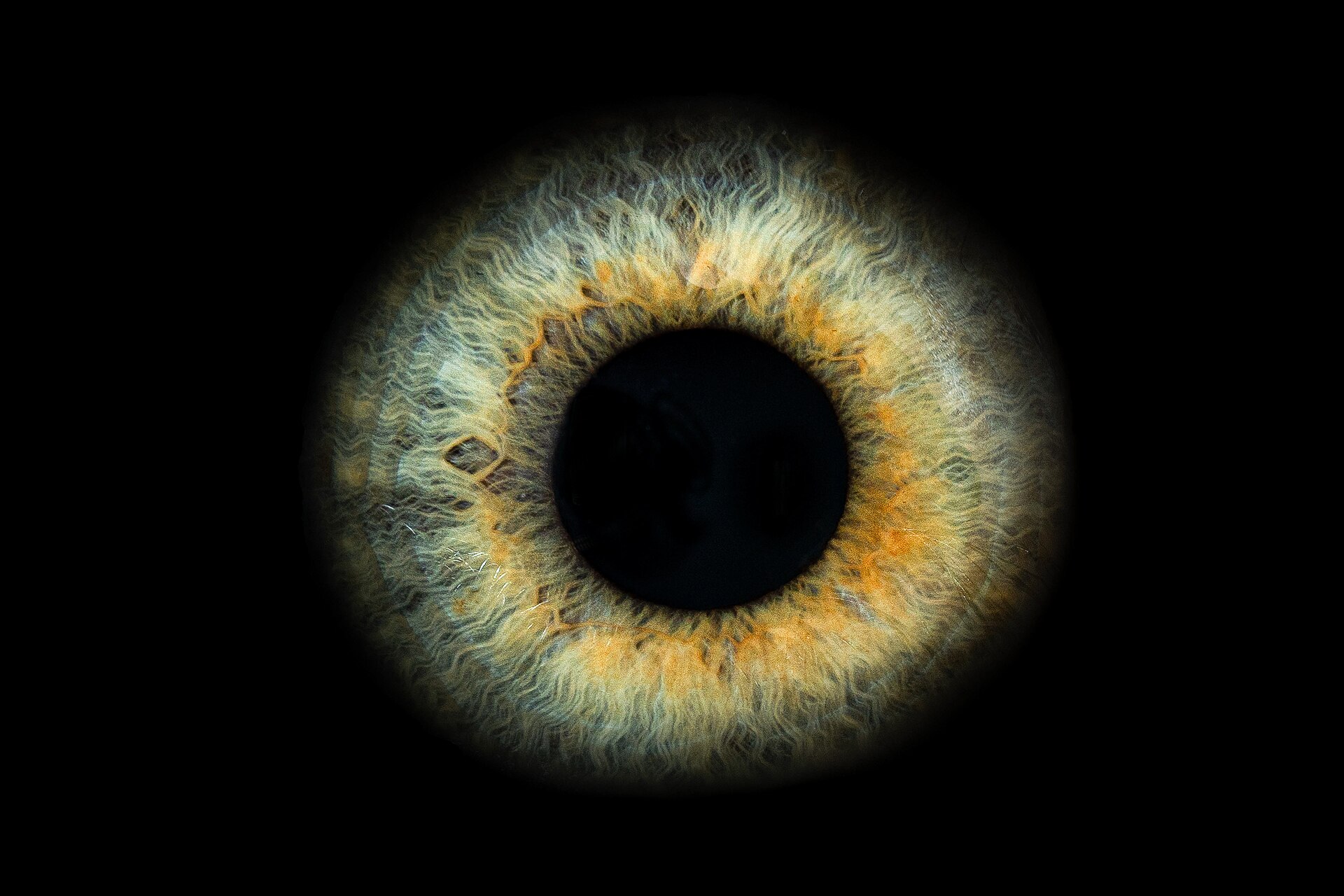
The human eye is the special sense organ of vision, and more than 50% of the body's sensory receptors are located in the eyes. In the adult, the eyeball measures about 2.5 centimeters in diameter, yet only about one-sixth of it is exposed to the outside air. Its sensitivity is confined to a narrow band of electromagnetic energy: the eye detects visible light at wavelengths from 400 to 700 nanometers.
1. Anatomy of the Eyeball
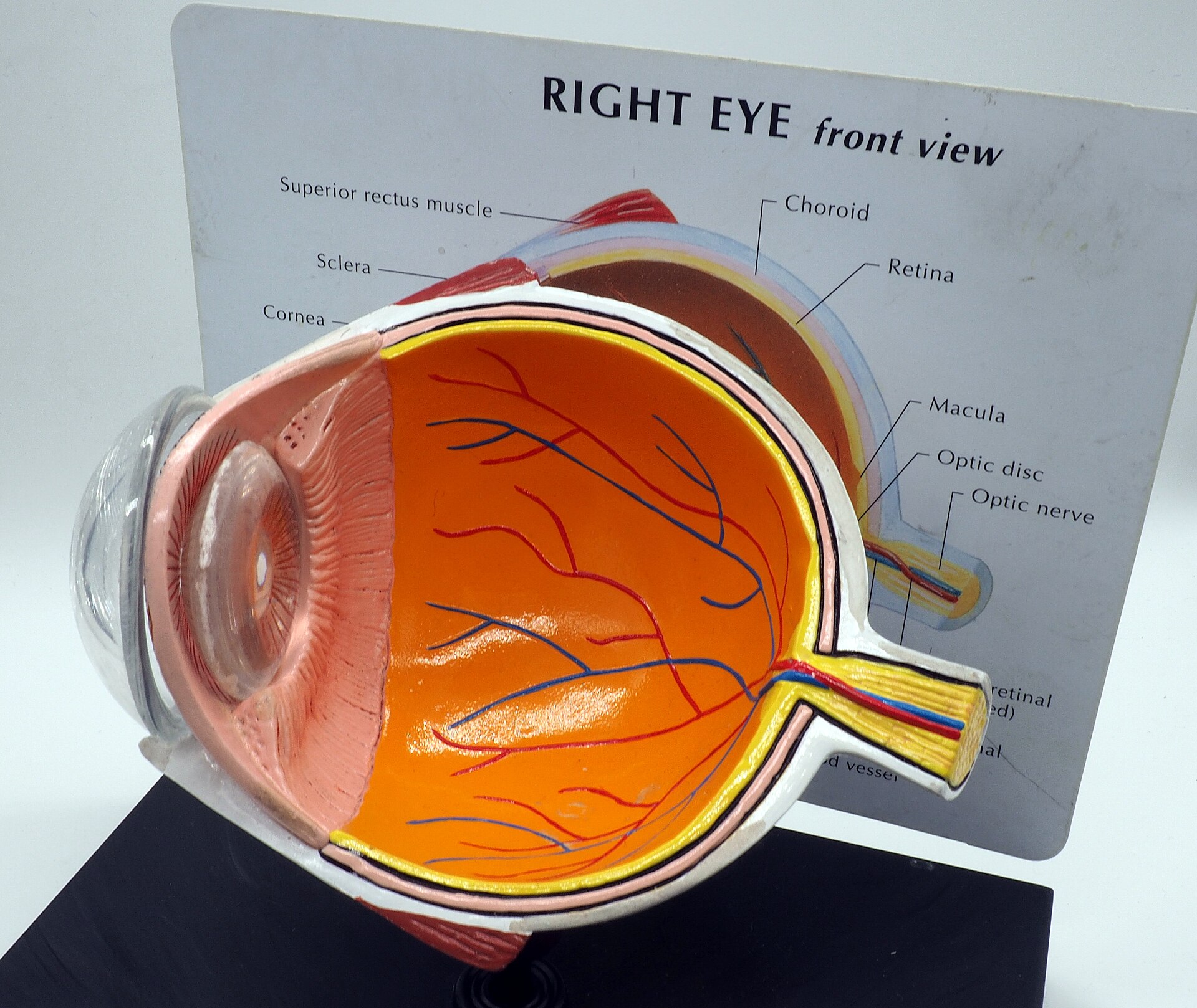
The wall of the eyeball is built from three concentric layers: the fibrous tunic, the vascular tunic, and the retina. The fibrous tunic forms the outermost coat and divides into the anterior cornea and the posterior sclera. The sclera is an opaque layer that surrounds the posterior five-sixths of the eyeball, while the cornea is a transparent layer continuous with it that occupies the anterior one-sixth. Functionally the most important of the three is the retina, which receives external visual stimuli.
The cornea itself is layered, with non-keratinized stratified squamous epithelium on its outer surface, collagen fibers and fibroblasts in its middle coat, and simple squamous epithelium on its inner surface. Its central region receives oxygen from the outside air. The corneal stroma accounts for around 90% of the thickness of the cornea.
2. Refractive Media and the Pathway of Light
The refractive media of the eye — the cornea, vitreous body, lens and aqueous humor — are the structures that help focus light onto the retina where it can be detected by the photoreceptors.Light first passes through the cornea, the clear dome-shaped front layer of the eye that bends light to help the eye focus, and then through the lens, a clear inner part of the eye that works with the cornea to focus light correctly on the retina.The iris, which is the colored part of the eye, controls how much light enters the eye through the pupil.The lens, a clear inner part of the eye just behind the pupil, changes shape to focus on objects that are close.
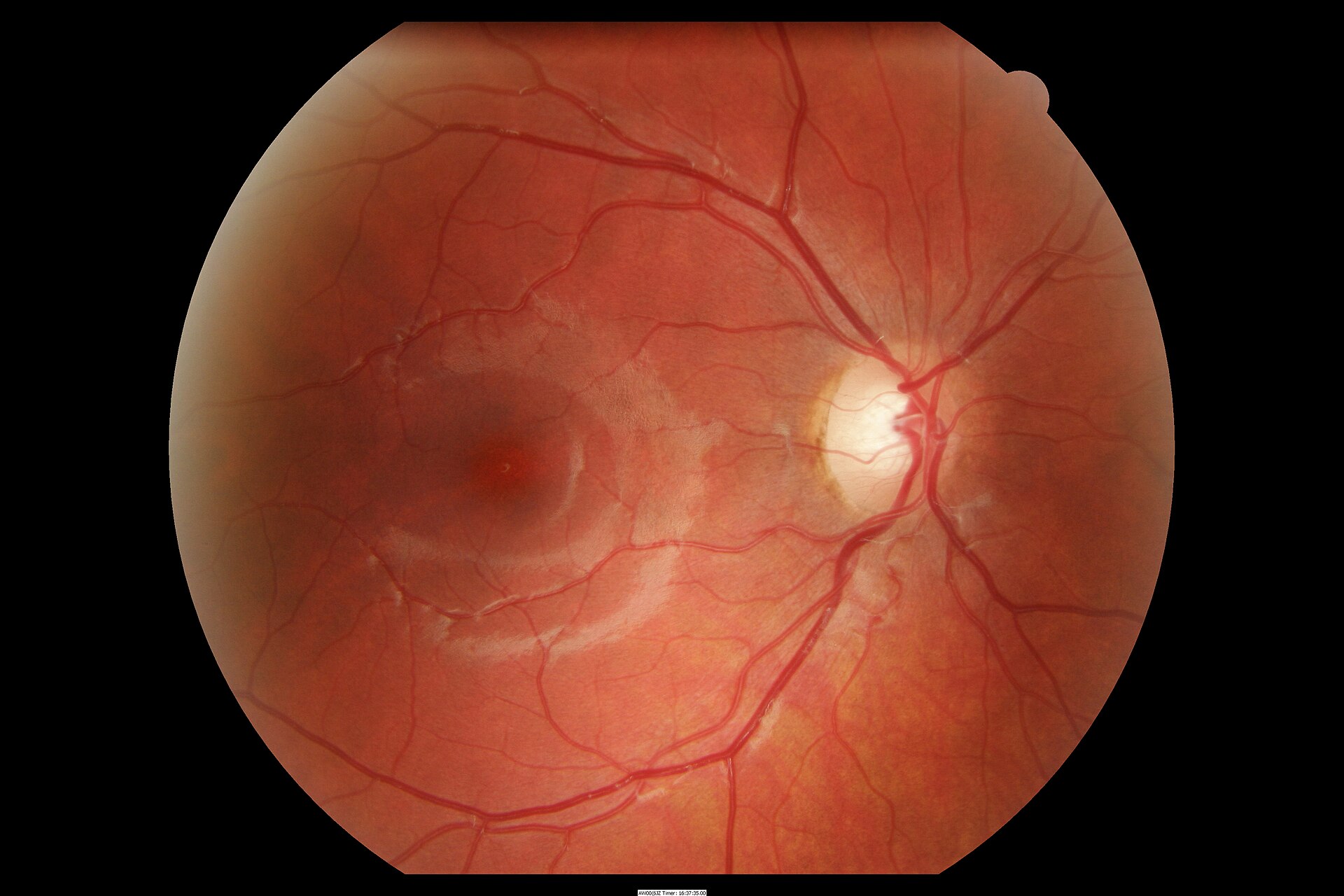
The anterior chamber holds aqueous humor, which the eye continually produces and drains through the drainage angle to keep eye pressure constant.Light focused by the cornea and lens passes through the vitreous onto the retina, the light-sensitive tissue lining the back of the eye, where the macula provides detailed central vision and the peripheral retina provides side vision.
3. The Retina and Photoreceptors
The retina is the light-sensitive tissue lining the back of the eye, and it is the nervous layer of the eyeball that receives external visual stimuli. The retina is composed of several layers and contains specialized cells for the initial processing of visual stimuli. The cells in the retina that respond to light stimuli are an example of a specialized receptor known as a photoreceptor, and photoreceptors are neurons that function as specialized receptor cells located in the retina of the eyeball, primarily responsible for the transduction of light stimuli for vision. The photoreceptors, rods and cones, change their membrane potential when stimulated by light energy, which alters the amount of neurotransmitter they release onto bipolar cells in the outer synaptic layer.
Each retina holds about 6 million cones and more than 100 million rods, giving a rod-to-cone ratio of about 20 to 1 except at the fovea centralis.Rod cells have elongated cylindrical outer segments resembling rods, with stacks of membrane-bound discs containing the photopigment rhodopsin, which consists of the protein scotopsin and the chromophore retinal; rods are strongly photosensitive and are located in the outer edges of the retina, where they detect dim light and serve primarily for peripheral and nighttime vision. Cones are weakly photosensitive and are located near the center of the retina, responding to bright light with a primary role in daytime color vision. Cones are important for visual acuity and contribute to high-resolution vision in bright conditions.
Rhodopsin reaches its peak sensitivity at 498 nanometers. The three cone types peak at 564 nm for long-wave cones, 534 nm for medium-wave cones and 420 nm for short-wave cones.Primates use a three-cone trichromatic system that results in full color vision, and the perceived color is a result of the ratio of activity of the three types of cones.
The fovea is a small area at the exact center of the retina that lacks supporting cells and blood vessels and contains only photoreceptors, which is why visual acuity is greatest there. Because the axons of retinal ganglion cells pass through the retina, there are no photoreceptors at the very back of the eye where the optic nerve begins, creating a blind spot in the retina and a corresponding blind spot in the visual field.
The shape change of retinal in the photoreceptors initiates visual transduction in the retina; activation of retinal and the opsin proteins results in activation of a G protein, which changes the membrane potential of the photoreceptor cell so that it releases less neurotransmitter into the outer synaptic layer.
4. Visual Pathway to the Brain
The visual pathway refers to the anatomical structures that convert light energy into electrical action potentials interpretable by the brain, beginning at the retina and terminating at the primary visual cortex.The optic nerves are formed by about one million myelinated axons arising from retinal ganglion cells that converge at the optic disc, and their fibers are myelinated by oligodendrocytes rather than Schwann cells. The optic chiasm is a point of union and decussation of the two optic nerves, where the nasal fibers of each eye cross the midline to join the temporal fibers of the contralateral eye.The lateral geniculate bodies are rounded protuberances on the inferior surface of the diencephalon that house the multi-layered lateral geniculate nuclei, whose six layers contain cells similar to those of the retinal ganglion layer.
5. Accessory Structures and Protection
The eye is protected from mechanical injury by being enclosed in a bony socket called the orbit, a four-sided pyramid whose floor is formed by parts of the maxilla, zygomatic, and palatine bones and whose roof includes the orbital plate of the frontal bone.The accessory structures of the eye include the eyelids, eyelashes, eyebrows, the lacrimal apparatus, and the extrinsic eye muscles, and the eyelids cover the eyes during sleep and spread lubricating secretions over the eyeball.
Six extraocular muscles attach to the sclera and move the eye up and down, from side to side, and in rotation. The conjunctiva is a mucous membrane that lines the lids and bends back over the surface of the eyeball, with a palpebral portion lining the lids and a bulbar portion covering the white of the eye.
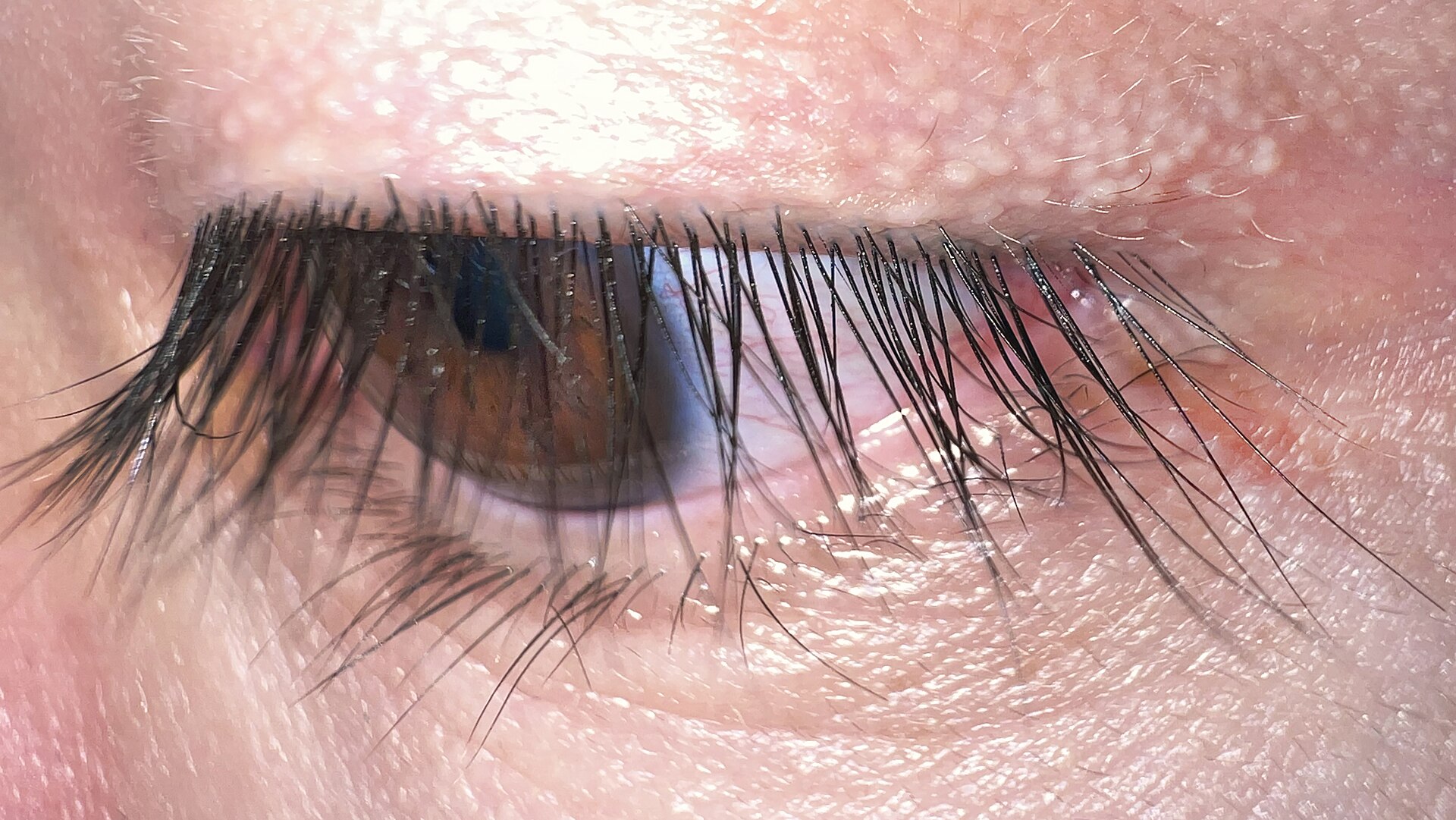
A three-layer tear film — mucous, watery and oily — keeps the front of the eye lubricated. The eye is kept moist by the secretions of the almond-shaped lacrimal glands, which lie under the upper lids and extend inward from the outer corner of each eye, with one portion resting in a depression of the frontal bone and the other projecting into the back of the upper lid.The ducts from each lacrimal gland, three to 12 in number, open into the superior conjunctival fornix, from which tears flow across the eye into the puncta lacrimalia at the margin of each eyelid and then through the lacrimal ducts into the lacrimal sacs and nasolacrimal ducts, which carry them into the nose.
Evaporation of the tears as they flow across the eye is largely prevented by oily and mucous secretions from other glands, including the meibomian, or tarsal, glands, which run through the tarsal plates and release an oil onto the lid margin that acts as a barrier for the tear fluid. The front surface of the eyeball, the cornea, must remain moist, which the eyelids achieve by sweeping the secretions of the lacrimal apparatus and other glands over it during waking hours and covering the eyes during sleep; the blink reflex also prevents injuries from foreign bodies.
6. Evolution of the Eye
Trilobite compound eyes stand as the earliest well-preserved visual surfaces in the fossil record, a status illustrated by Telephina convexa, an animal that swam in the oceans around 450 million years ago. A different form of early vision belonged to Anomalocaris, an early arthropod relative whose giant compound eyes were set on the ends of stalks and built from repeating units known as ommatidia.
Nilsson's calculations assumed a tiny change with each generation towards a more evolved eye. Even under that model, his pessimistic estimate of the time required for an eye to evolve was less than 364,000 years.
7. Historical Milestones in Eye Science
-
1851Helmholtz's ophthalmoscope and ophthalmometerHelmholtz introduces the ophthalmoscope and the ophthalmometer, also known as the keratometer.
-
7 December 1905First successful penetrating keratoplasty in a humanEduard Zirm performs the first successful penetrating keratoplasty in a human in Olmutz near Prague, on Alois Glogar, a 45-year-old farm labourer who had sustained severe bilateral alkali burns 16 months earlier while cleaning his chicken coop with lime. Donor tissue came from an 11-year-old boy blind from a penetrating scleral injury; two 5-mm buttons were removed by von Hippel trephine, and bilateral transplants were carried out under chloroform anaesthesia with strict asepsis. 10Centennial review of corneal transplantation · Clinical & Experimental Ophthalmology (Wiley); hosted by University of Iowa ↗
-
1911Nobel Prize for work on the dioptrics of the eyeAllvar Gullstrand receives the Nobel Prize in Physiology or Medicine for work on the dioptrics of the eye.
-
1967Nobel Prize for visual processesThe Nobel Prize in Physiology or Medicine is awarded jointly to Ragnar Granit, Haldan Keffer Hartline and George Wald for their discoveries concerning the primary physiological and chemical visual processes in the eye. 11The Nobel Prize in Physiology or Medicine 1967 — Granit, Hartline and Wald · Nobel Prize Outreach ↗
-
1981Nobel Prize for visual information processingThe Nobel Prize in Physiology or Medicine is divided, one half going jointly to David H. Hubel and Torsten N. Wiesel for their discoveries concerning information processing in the visual system. 12The Nobel Prize in Physiology or Medicine 1981 — Sperry, Hubel and Wiesel · Nobel Prize Outreach ↗
8. Clinical Significance and Global Burden
Unoperated cataract accounts for a higher proportion of vision impairment in low- and middle-income countries than in high-income countries, where glaucoma and age-related macular degeneration are more common. At least 1 billion of those cases could have been prevented or have yet to be addressed.
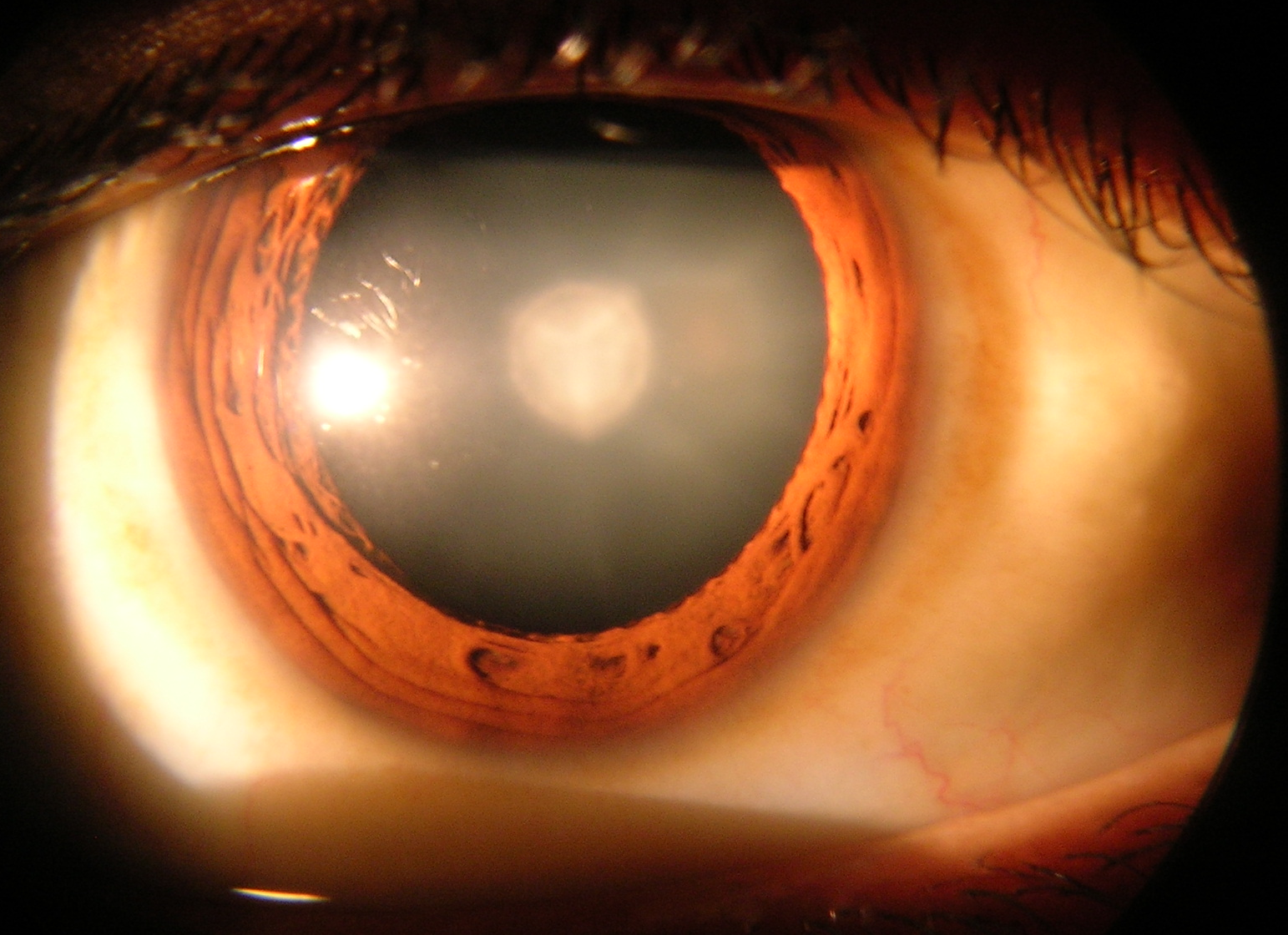
Over time cataracts can make vision blurry, hazy, or less colorful, interfere with reading and other everyday activities, and eventually lead to vision loss. Cataract surgery removes the clouded lens and replaces it with an artificial intraocular lens; 9 out of 10 people who have the surgery can see better afterwards.
The most common type of color vision deficiency makes it hard to tell red from green, another type makes blue and yellow look the same, and rare cases involve seeing no color at all.
9. Vision Data and Surveillance
Eye health data are reported as estimates, and they may show a higher or lower number of eye disease cases than a population actually has, depending on how researchers collected the data. Department of Health and Human Services leads the Healthy People 2030 national public health initiative, and the National Eye Institute leads its vision workgroup.
The CDC's Vision and Eye Health Data Portal provides detailed datasets on eye health, while the National Vision and Eye Health Surveillance System shares estimates on vision loss and blindness in the United States.
10. The Eye in Spaceflight
Prolonged weightlessness allows bodily fluids, including blood and cerebrospinal fluid, to shift toward the head, a redistribution that can produce swelling of the optic nerve and folds in the retina. The cluster of eye and brain changes observed in astronauts is designated spaceflight associated neuro-ocular syndrome, or SANS. The likelihood of developing SANS rises with the length of a space mission, and the condition remains a leading research priority for scientists preparing for a journey to Mars.
11. Timeline: from the first eyes to 2030 eye-care targets
- 545 to 530 million years agoThe Cambrian explosion and the first eyesThe dramatic increase in animal diversity between 545 and 530 million years ago is known as the Cambrian explosion; the evolution of the eye is thought to have been a catalyst for the arms race between increasingly aware organisms that followed.
- about 514 million years agoThe giant compound eyes of AnomalocarisFossils from South Australia preserved the eye surface of the early arthropod relative Anomalocaris, whose giant compound eyes sat on stalks and were studied in three dimensions for the first time in 2011.
- about 450 million years agoTrilobite eyes enter the fossil recordTrilobite compound eyes are the earliest well-preserved visual surfaces in the fossil record, and the trilobite Telephina convexa swam in the oceans around 450 million years ago.
- 1760The idea of cutting out an opaque corneaErasmus Darwin, grandfather of Charles Darwin, first suggested the removal of an opaque cornea with a small trephine, proposing that an ingenious surgeon or oculist should try it.
- 1789The first monograph on ophthalmic surgeryGuillaume Pellier de Quengsy, a distinguished cataract surgeon, published the first monograph devoted to ophthalmic surgery.
- 1824Animal corneas proposed as graftsFranz Riesinger was the first to propose replacing opaque human corneas with transparent animal corneas, after witnessing an attempt at skin grafting in England.
- 1851Helmholtz invents the ophthalmoscopeAmong Helmholtz's most valuable inventions were the ophthalmoscope and the ophthalmometer, both made in 1851; the ophthalmoscope remains one of the most important instruments of the physician.
- 7 December 1905First successful human corneal transplantIn the small town of Olmutz near Prague, Eduard Zirm performed the first successful penetrating keratoplasty in a human in which the graft remained clear, using tissue from an 11-year-old donor.
- 1911Nobel Prize for the dioptrics of the eyeAllvar Gullstrand received the Nobel Prize in Physiology or Medicine for his work on the dioptrics of the eye.
- 1967Nobel Prize for the visual processes in the eyeThe prize was awarded jointly to Ragnar Granit, Haldan Keffer Hartline and George Wald for their discoveries concerning the primary physiological and chemical visual processes in the eye.
- 1981Nobel Prize for information processing in the visual systemOne half of the prize went jointly to David H. Hubel and Torsten N. Wiesel for their discoveries concerning information processing in the visual system.
- 1994A model of how fast an eye can evolveDan-Eric Nilsson of Lund University modelled the evolution of a simple layer of light receptors into an eye capable of seeing high-resolution images, with a pessimistic estimate of less than 364,000 years for the change.
- 2011First approved retinal prosthesisThe Argus II retinal prosthesis, or bionic eye, became the first approved solution for retinitis pigmentosa, though it was only available in a handful of countries.
- 2019The WHO World report on visionWHO's work on eye care is guided by the recommendations of the World report on vision and by the resolution on integrated, people-centred eye care adopted at the Seventy-third World Health Assembly in 2020.
- 2030Global targets for integrated eye careWHO works with Member States to monitor the global targets for 2030 on integrated people-centred eye care, including the SPECS 2030 initiative for the more than 800 million people in need of refractive error services.
12. Frequently asked questions
What is the eye's function among the body's sensory receptors?
It is the special sense organ of vision, and more than 50% of the body's sensory receptors are located in the eyes 1Anatomy, Head and Neck, Eye · StatPearls via NCBI Bookshelf, U.S. National Library of Medicine ↗.
Which wavelengths of visible light can the eye detect?
It detects wavelengths from 400 to 700 nanometers 1.
How many photoreceptors does each retina contain?
Each retina holds about 6 million cones and more than 100 million rods 1.
How many refractive media does the eye have, and what are they?
There are four: the cornea, aqueous humor, lens and vitreous body 2Eyeball: Structure and function · Kenhub ↗.
How many people worldwide live with vision impairment, and how many of those cases are preventable or unaddressed?
At least 2.2 billion people live with a near or distance vision impairment, and at least 1 billion of those cases could have been prevented or have yet to be addressed 13Blindness and vision impairment · World Health Organization ↗.
What happens to astronauts' eyes during spaceflight?
About 70 percent of astronauts aboard the International Space Station experience swelling in the back of their eyes 17Risk of Spaceflight Associated Neuro-ocular Syndrome (SANS) · National Aeronautics and Space Administration (NASA) ↗.
References (19)
- 1Anatomy, Head and Neck, Eye — StatPearls via NCBI Bookshelf, U.S. National Library of Medicine Web↩View source
- 2Eyeball: Structure and function — Kenhub Web↩View source
- 3Eye Anatomy: Parts of the Eye and How We See — American Academy of Ophthalmology WebView source
- 4Anatomy and Physiology 2e — 14.1 Sensory Perception — OpenStax, Rice University WebView source
- 5Biology 2e — 36.5 Vision — OpenStax, Rice University WebView source
- 6Photoreceptors: Rods and cones — Kenhub WebView source
- 7The visual pathway: Anatomy, components and histology — Kenhub WebView source
- 8Human eye — Encyclopaedia Britannica WebView source
- 9Eyes on the prize: the evolution of vision — Natural History Museum, London WebView source
- 10Centennial review of corneal transplantation — Clinical & Experimental Ophthalmology (Wiley); hosted by University of Iowa 2023 Book↩View source
- 11The Nobel Prize in Physiology or Medicine 1967 — Granit, Hartline and Wald — Nobel Prize Outreach 1967 Web↩View source
- 12The Nobel Prize in Physiology or Medicine 1981 — Sperry, Hubel and Wiesel — Nobel Prize Outreach 1981 Web↩View source
- 13Blindness and vision impairment — World Health Organization Web↩View source
- 14Cataracts — National Eye Institute (NEI), U.S. National Institutes of Health WebView source
- 15Color Blindness — National Eye Institute (NEI), U.S. National Institutes of Health WebView source
- 16Eye Health Data and Statistics — National Eye Institute (NEI), U.S. National Institutes of Health WebView source
- 17Risk of Spaceflight Associated Neuro-ocular Syndrome (SANS) — National Aeronautics and Space Administration (NASA) Web↩View source
- 18Hermann von Helmholtz — Encyclopaedia Britannica WebView source
- 19The Nobel Prize in Physiology or Medicine 1911 — Allvar Gullstrand — Nobel Prize Outreach 1911 WebView source