Microscope
The instrument that opened the invisible world - from Dutch spectacle lenses and Hooke's cork cells to electron, confocal and cryo-electron microscopy.
The microscope is an instrument that produces enlarged images of small objects, revealing details too small to be seen by the unaided eye. From compound microscopes credited to Dutch spectacle makers about 1590, Robert Hooke's Micrographia and Antonie van Leeuwenhoek's single-lens observations of bacteria and protozoa, it became the foundational tool of biology and medicine. In the twentieth century electron microscopy broke the resolution limit of light, while phase contrast, confocal, super-resolution fluorescence and cryo-electron microscopy extended imaging to living cells, single molecules and atomic detail.
In this article
- 1Overview
- 2Optical Microscopes: Simple and Compound
- 3Origins and Early Development
- 4Hooke, Leeuwenhoek, and the Microscopic World
- 5Lens Theory and the Resolution Limit
- 6The Electron Microscope
- 7Confocal and Super-Resolution Microscopy
- 8Applications and Impact
- 9Timeline of Key Developments
- 10Key Figures
- 11Timeline: from the first compound microscopes to cryo-electron microscopy
- 12Knowledge graph
Overview
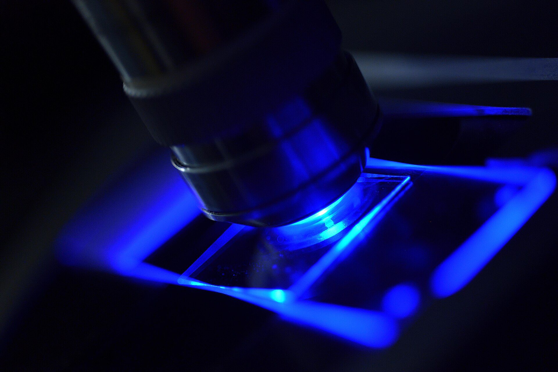
A microscope is an instrument that makes an enlarged image of a small object, revealing details too small to be seen by the unaided eye; the most familiar type is the optical microscope, which uses visible light focused through lenses. The word microscope comes from the Latin microscopium, itself derived from the Greek mikros, meaning small, and skopein, meaning to look at.
1. Optical Microscopes: Simple and Compound
Optical microscopes fall into two broad classes: the simple microscope, which relies on a single lens, and the compound microscope, in which several optical components are arranged in line. A simple microscope can resolve below 1 micrometre, while a compound microscope can resolve down to about 0.2 micrometres. The best lenses made by Antonie van Leeuwenhoek achieved magnifying powers of 50 to 300 times, and it was with single-lens instruments of his own making that he produced the first observations of bacteria and protozoa. Leeuwenhoek was arguably the first person to bring the light microscope properly to the attention of natural scientists interested in the study of living things.
The optical train of a microscope is rarely perfect: such systems typically suffered from as many as five common aberrations — spherical, chromatic, curvature of field, comatic, and astigmatic. Chromatic aberration was the particular weakness of the compound instruments of Leeuwenhoek's era, which is why his single high-quality lenses of very short focal length were then preferable. Because of diffraction, the image of a point source is not a point but an Airy pattern, a central spot known as the Airy disk surrounded by multiple diffraction rings. Working distances on modern microscopes can range from 3 to 5 centimetres and as much as 20 centimetres in some models, leaving considerable room between the objective and the specimen.
Contrast techniques extend what the optical microscope can reveal. In bright field microscopy, light absorption is responsible for image formation, whereas phase contrast microscopy converts phase differences into visible contrast. The phase contrast microscope became particularly important in the study of living cells. The twentieth century brought a renaissance in microscope technology through new contrast-enhancing techniques such as phase contrast, Hoffman modulation contrast, differential interference contrast, and fluorescence microscopy.
2. Origins and Early Development
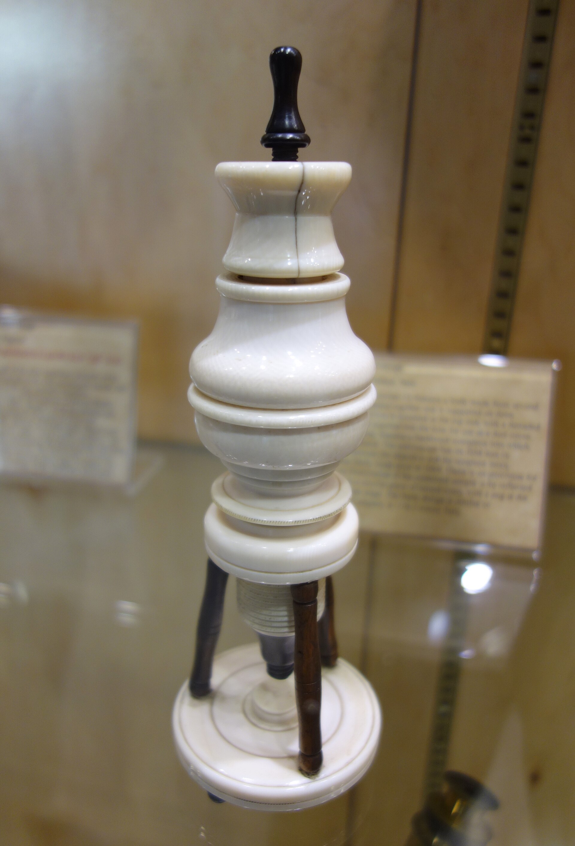
The compound microscope is generally credited to Dutch spectacle makers — Hans Jansen, his son Zacharias Jansen, and Hans Lippershey — and dated to about 1590 in the Netherlands. The first recorded microscope was built by Zacharias and Hans Janssen in the 1590s. In its early form the instrument consisted of hollow metal tubes with a lens on each end, a modest arrangement from which the modern super-resolution microscope would eventually descend.
3. Hooke, Leeuwenhoek, and the Microscopic World
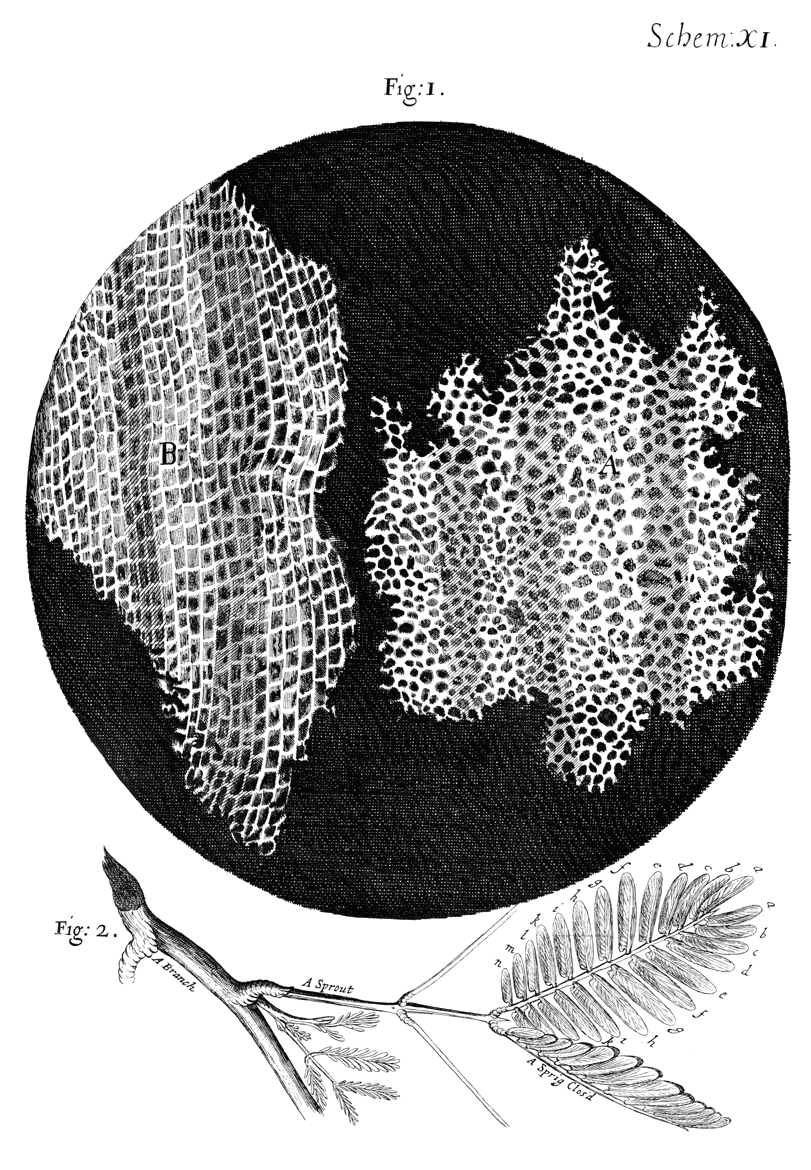
Robert Hooke was appointed curator of experiments to the Royal Society of London in 1662 and was elected a fellow the following year. In 1665 he published what would become his most famous work, Micrographia, or Small Drawings. Examining an exceedingly thin piece of cork, he could exceeding plainly perceive it to be all perforated and porous, much like a honeycomb, though the pores of it were not regular. He observed that these pores, or cells, were not very deep but consisted of a great many little boxes separated out of one continued long pore by certain diaphragms. Hooke was born on July 18, 1635, at Freshwater, Isle of Wight, England, and died on March 3, 1703, in London.
Antonie van Leeuwenhoek was born on October 24, 1632, in Delft, and died on August 26, 1723, in Delft. As the story goes, he was inspired by Robert Hooke's Micrographia of 1665 and went on to refine his own methods of producing lenses. In turn, the Royal Society published a large part of his correspondence in the Philosophical Transactions and made him a Fellow.
4. Lens Theory and the Resolution Limit
Around 1830 Joseph Jackson Lister, working with the instrument maker William Tulley, produced one of the first microscopes to correct for both spherical and chromatic aberration. In 1868 Ernst Abbe introduced an apochromatic system of lenses whose colour correction surpassed that of achromatic lenses, and in 1873 he set out a comprehensive analysis of lens theory.
For a long time optical microscopy was held back by a presumed limitation: that it would never obtain a better resolution than half the wavelength of light.
5. The Electron Microscope
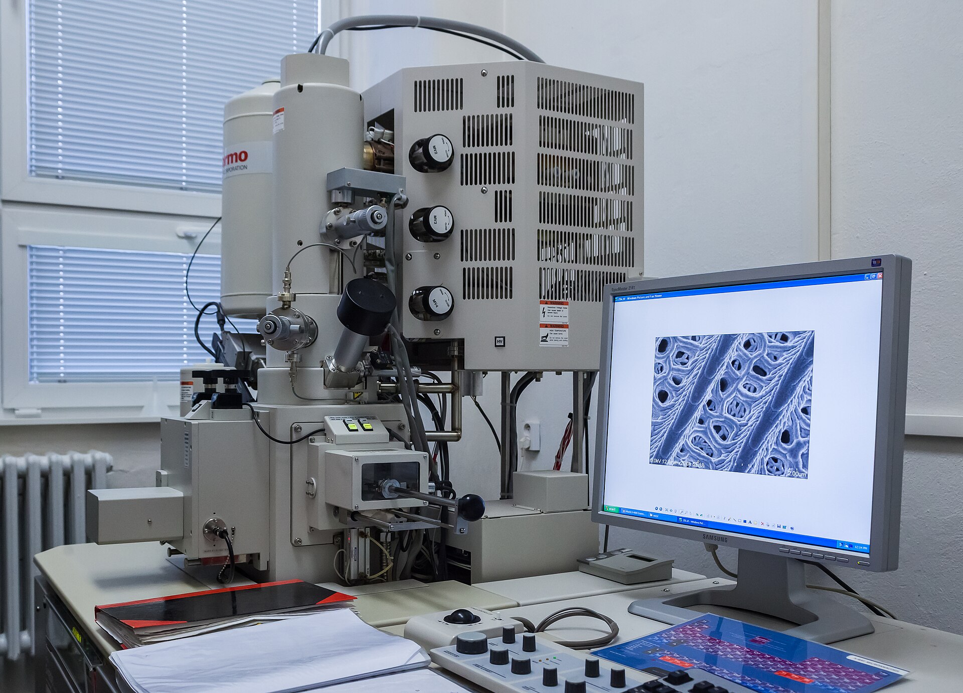
An electron microscope is a microscope that attains extremely high resolution using an electron beam instead of a beam of light to illuminate the object of study. Because the wavelength of electrons is much shorter than that of light, the electron microscope can make very small structures visible, even the position of individual atoms. De Broglie derived a formula for electron wavelength showing that electrons accelerated by 60 kilovolts would have an effective wavelength of 0.05 angstrom, about one hundred-thousandth that of green light. In 1926 it was demonstrated that magnetic or electrostatic fields could serve as lenses for electrons, and by 1931 the German electrical engineers Max Knoll and Ernst Ruska had devised a two-lens electron microscope that produced images of the electron source. In 1933 a primitive electron microscope was built that imaged a specimen rather than the electron source, and in 1935 Knoll produced a scanned image of a solid surface. The conventional electron microscope requires that the electron beam be in a vacuum, because electrons cannot ordinarily travel an appreciable distance in air at atmospheric pressure. In practice, electron microscope resolutions down to about 1 angstrom have been achieved, close to the theoretical limit set by quantum mechanics; the transmission electron microscope can magnify more than 1,000,000 times.
Ernst Ruska was born December 25, 1906 in Heidelberg and died May 27, 1988 in West Berlin. He discovered that a magnetic coil could be used as a lens for electron beams and in 1933 developed the first electron microscope, having very quickly improved various details so that it had a performance clearly superior to that of the conventional light microscope. In Berlin-Spandau in 1937 he and Bodo von Borries set up the Laboratory for Electron Optics and developed there until 1939 the first customised electron microscopes, the Siemens Super Microscope; in 1939 the first commercially available, mass-produced electron microscope entered the market after Ruska was appointed by Siemens to take part in its development. One half of the 1986 Nobel Prize in Physics was awarded to him for his fundamental work in electron optics and for the design of the first electron microscope.
In the scanning electron microscope a beam of electrons is scanned over the surface of a solid object to build up an image of the details of the surface structure; its advantage is its large depth of focus, which gives a three-dimensional image as opposed to the sectional image obtained with a transmission microscope. The earliest known work describing the concept was by M. Knoll in 1935, who along with other pioneers in electron optics was working in Germany. Von Ardenne constructed a scanning transmission electron microscope in 1938 by adding scan coils to a transmission electron microscope, and the first scanning electron microscope with a sub-micron probe was developed by von Ardenne, a private consultant who had his own laboratory in Berlin, over a period of about two years. The SEM work was taken up by Dennis McMullan in 1948 and he and Charles Oatley built their first scanning electron microscope; by 1952 the Cambridge SEM had achieved a resolution of 50 nm and produced the first micrographs showing the striking three-dimensional imaging characteristics of the modern-day SEM. The beginning of the general use of the SEM can be accurately dated to 1965 when the Cambridge Instrument Company marketed their Stereoscan 1 SEM, and scanning electron microscopes number in excess of 50,000 world-wide.
6. Confocal and Super-Resolution Microscopy
The confocal approach rests on spatial filtering: light that originates away from the plane of focus is rejected rather than collected, which matters most in specimens thicker than that plane. Marvin Minsky patented the idea of rejecting out-of-focus light in the 1950s, and realized it through pinhole apertures placed on both the illumination and the detection sides in the same conjugate image plane — the arrangement that makes the two apertures confocal. The detection-side pinhole barred light coming from above or below the plane of focus from reaching the photomultiplier. Sketches of confocal beam paths using a pinhole appear in papers from the 1940s and early 1950s, but the first prototype confocal microscope was invented, patented and built by Minsky in 1955/56, growing out of his search for a better way to image unstained neural tissue and first produced in 1955 while he was at Harvard University.
A modern confocal microscope keeps the pinholes, objective lenses and low-noise detectors of the original design, and typically adds fast scanning mirrors, filters for wavelength selection, and laser illumination. Closing the pinholes to their minimum size yields a diffraction-limited imaging system, improving lateral resolution over a conventional widefield fluorescence microscope. In 1987 White and Amos were the first to build a confocal laser scanning microscope in which the laser beam itself performed the scanning rather than a moving stage, a change that significantly sped up imaging.
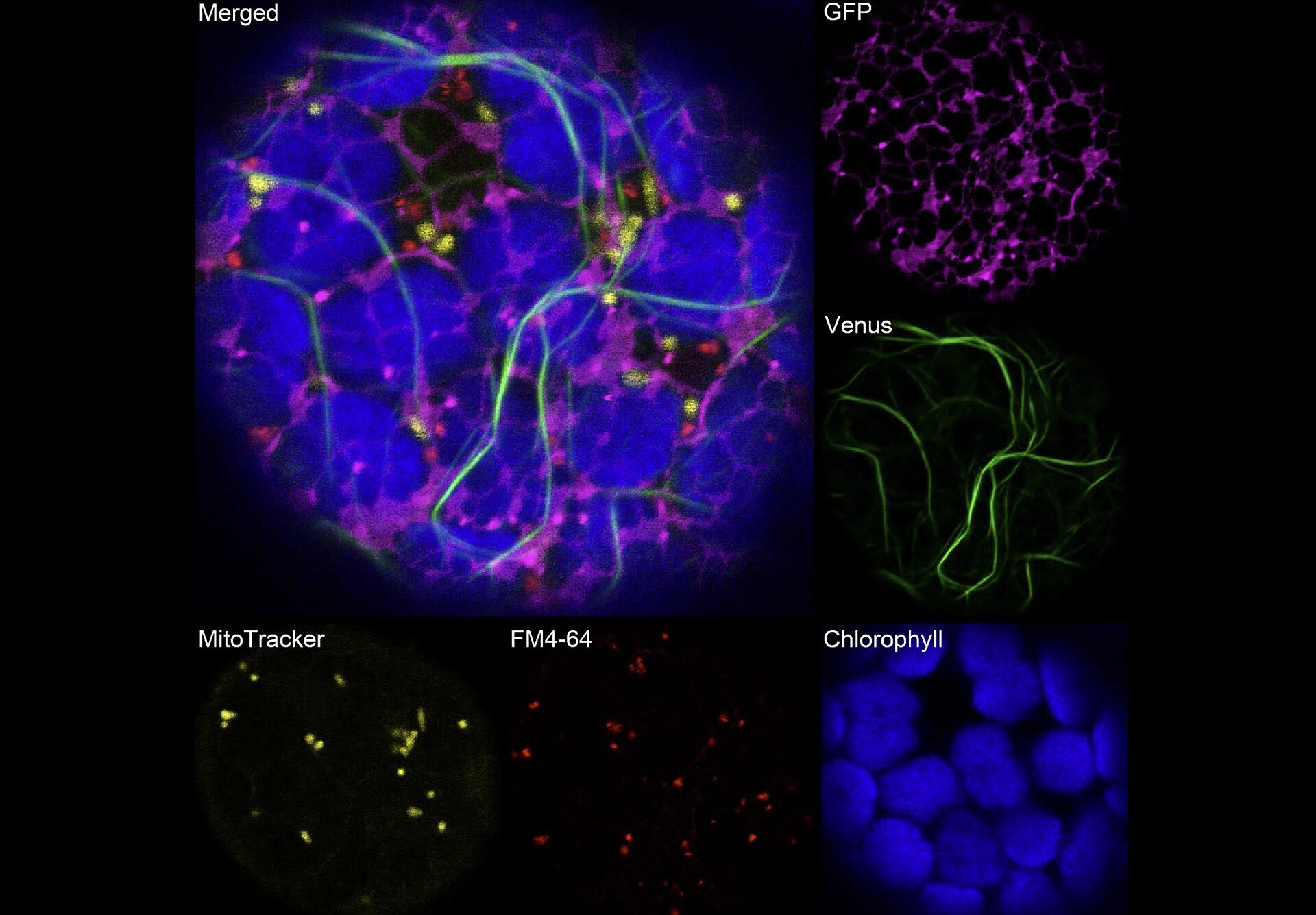
Stimulated emission depletion microscopy, developed by Stefan Hell in 2000, scans the sample nanometre by nanometre to produce an image whose resolution surpasses Abbe's stipulated limit. Cryo-electron microscopy, honoured with the Nobel Prize in Chemistry 2017 awarded to Jacques Dubochet, Joachim Frank and Richard Henderson, both simplifies and improves the imaging of biomolecules. Frits Zernike developed a way to make phase differences in light visible in the early 1930s; and he received the 1953 Nobel Prize in Physics for it.
7. Applications and Impact
Over about 300 years the light microscope evolved from a simple single lens device into an instrument capable of observing the dynamics of single biological molecules inside living cells and tracking every cell nucleus in the development of whole embryos and plants.
Nearly 200 years later in 1838 Matthias Schleiden, a German botanist who made extensive microscopic observations of plant tissues, described them as being composed of cells. Rudolf Virchow popularized the concept of cell theory in an essay entitled Cellular Pathology, using the Latin phrase omnis cellula a cellula, meaning all cells arise from cells, which is essentially the second tenet of modern cell theory.
8. Timeline of Key Developments
-
1926Fields as electron lensesMagnetic and electrostatic fields demonstrated as electron lenses.17Electron microscope | Uses, Advantages & Limitations · Encyclopaedia Britannica ↗
-
1931Two-lens electron microscopeKnoll and Ruska's two-lens design.17
-
1937Laboratory for Electron OpticsIn Berlin-Spandau, Ruska and Bodo von Borries set up the Laboratory for Electron Optics.18Ernst Ruska - Biographical · Nobel Prize Outreach (Royal Swedish Academy of Sciences) ↗
-
1939Siemens Super MicroscopeThe laboratory developed the first customised electron microscopes, the Siemens Super Microscope.18
-
Around 1830Correction of aberrationJoseph Jackson Lister, in collaboration with instrument maker William Tulley, made one of the first microscopes that corrected for both spherical and chromatic aberration.12The Microscope · Science Museum Group, London ↗
9. Key Figures
| Name | Born | Died | Principal contribution |
|---|---|---|---|
| Robert Hooke | July 18, 1635, Freshwater, Isle of Wight, England | March 3, 1703, London | Micrographia, first use of the word cell |
| Antonie van Leeuwenhoek | October 24, 1632, Delft | August 26, 1723, Delft | self-taught lens maker |
| Ernst Ruska | December 25, 1906, Heidelberg | May 27, 1988, West Berlin | designed the first electron microscope in 1933 |
10. Timeline: from the first compound microscopes to cryo-electron microscopy
- about 1590The compound microscope emergesDutch spectacle makers Hans Jansen, his son Zacharias Jansen and Hans Lippershey are credited with inventing the compound microscope, an instrument of an eyepiece and an objective lens that became increasingly popular in the mid-17th century.
- 1665Hooke publishes Micrographia and names the cellRobert Hooke published Micrographia, a folio volume of microscopic views of familiar objects, and in it first used the word cell for the microscopic honeycomb cavities he saw in cork.
- 1674Leeuwenhoek launches microbiologyUsing a single-lens microscope of his own making, Antonie van Leeuwenhoek observed protozoa for the first time; the work effectively launched microbiology.
- 1683First image of bacteriaThe first representation of bacteria appears in a drawing by Leeuwenhoek published in the Royal Society's Philosophical Transactions.
- 1838Schleiden describes plant tissues as cellsMatthias Schleiden, a German botanist who made extensive microscopic observations of plant tissues, described them as being composed of cells; Theodor Schwann reached the same conclusion for animal tissue the next year.
- 1855Virchow popularizes cell theoryRudolf Virchow published an essay entitled Cellular Pathology that popularized the concept omnis cellula a cellula, all cells arise from cells, today the second tenet of modern cell theory.
- 1873Abbe states the optical resolution limitErnst Abbe stipulated a physical limit for the maximum resolution of traditional optical microscopy: it could never become better than 0.2 micrometres.
- 1931Knoll and Ruska build a two-lens electron microscopeAfter magnetic and electrostatic fields were shown in 1926 to act as lenses for electrons, Max Knoll and Ernst Ruska devised a two-lens electron microscope that produced images of the electron source.
- 1933First electron microscope superior to light opticsErnst Ruska built the first electron microscope whose performance was clearly superior to that of the conventional light microscope.
- 1953Zernike wins the Nobel Prize for phase contrastFrits Zernike, who had developed a way to make phase differences in light visible in the early 1930s, received the Nobel Prize in Physics; the phase contrast microscope became particularly important in the study of living cells.
- 1955Minsky builds the first confocal microscopeSeeking a way to image unstained neural tissue, Marvin Minsky produced the first confocal scanning microscope, using pinholes to reject out-of-focus light.
- 1965The SEM reaches the marketThe Cambridge Instrument Company marketed the Stereoscan 1, the first commercial scanning electron microscope, followed about six months later by JEOL in Japan.
- 1986Nobel Prize for the electron microscopeErnst Ruska received one half of the Nobel Prize in Physics for his fundamental work in electron optics and for the design of the first electron microscope; Gerd Binnig and Heinrich Rohrer shared the other half for the scanning tunnelling microscope.
- 2013Cryo-EM reaches atomic resolutionWith a new type of electron detector, the final technical hurdle of cryo-electron microscopy was overcome and the desired atomic resolution was reached.
- 2014Nobel Prize for super-resolved fluorescence microscopyEric Betzig, Stefan W. Hell and William E. Moerner shared the Nobel Prize in Chemistry for developing methods that bypassed Abbe's limit, bringing optical microscopy into the nanodimension.
- 2017Nobel Prize for cryo-electron microscopyJacques Dubochet, Joachim Frank and Richard Henderson received the Nobel Prize in Chemistry for the development of cryo-electron microscopy, which both simplifies and improves the imaging of biomolecules.
References (23)
- 1Microscope | Types, Parts, History, Diagram, & Facts — Encyclopaedia Britannica WebView source
- 2Antonie van Leeuwenhoek | Biography, Discoveries, & Facts — Encyclopaedia Britannica WebView source
- 3From Animaculum to single molecules: 300 years of the light microscope — Open Biology, via PubMed Central (NCBI) WebView source
- 4Molecular Expressions Microscopy Primer: Anatomy of the Microscope — Florida State University / National High Magnetic Field Laboratory WebView source
- 5Microscopy - Medical Imaging Systems — NCBI Bookshelf (National Library of Medicine) WebView source
- 6Molecular Expressions Microscopy Primer: Museum of Microscopy — Florida State University / National High Magnetic Field Laboratory WebView source
- 7Frits Zernike - Facts — Nobel Prize Outreach (Royal Swedish Academy of Sciences) 1953 WebView source
- 8Microscope - Optics, Magnification, Invention — Encyclopaedia Britannica WebView source
- 9Robert Hooke | Biography, Discoveries, & Facts — Encyclopaedia Britannica WebView source
- 10Micrographia, by Robert Hooke — Project Gutenberg WebView source
- 11Focus on Leeuwenhoek — The Royal Society 2023 WebView source
- 12The Microscope — Science Museum Group, London Web↩View source
- 13Press release: The Nobel Prize in Chemistry 2014 — Nobel Prize Outreach (Royal Swedish Academy of Sciences) 2014 WebView source
- 14Confocal Microscopy: Principles and Modern Practices — PubMed Central (NCBI) WebView source
- 15Press release: The 2017 Nobel Prize in Chemistry — Nobel Prize Outreach (Royal Swedish Academy of Sciences) 2017 WebView source
- 16Foundations of Modern Cell Theory (Microbiology 3.2) — OpenStax, Rice University WebView source
- 17Electron microscope | Uses, Advantages & Limitations — Encyclopaedia Britannica Web↩View source
- 18Ernst Ruska - Biographical — Nobel Prize Outreach (Royal Swedish Academy of Sciences) 1986 Web↩View source
- 19Press release: The 1986 Nobel Prize in Physics — Nobel Prize Outreach (Royal Swedish Academy of Sciences) 1986 WebView source
- 20Ernst Ruska - Facts — Nobel Prize Outreach (Royal Swedish Academy of Sciences) 1986 WebView source
- 21Molecular Expressions: Marvin Lee Minsky — Florida State University / National High Magnetic Field Laboratory WebView source
- 22Scanning Electron Microscopy 1928-1965 (D. McMullan) — University of Cambridge, Department of Engineering 1928 BookView source
- 23The Early History and Development of The Scanning Electron Microscope — University of Cambridge, Department of Engineering BookView source
 Isaac Newton
 SpaceX
 Alan Turing: Computability, Bletchley Park, and the Turing Test
